Bio-Imaging
Contact
Dr Bob Asselbergh, ORCID
e-mail: Bob.Asselbergh@uantwerpen.vib.be
telephone: +32 3 265 1034
Simona Manzella
e-mail: Simona.manzella@uantwerpen.vib.be
telephone: +32 3 265 1917
location: CDE V.A.0.21
Mission
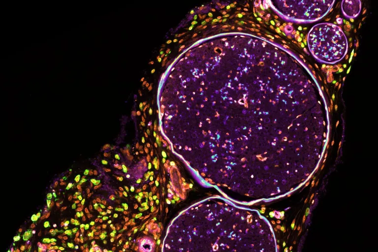
Bio-imaging is a principal field on the road to scientific discovery and is crucial to answer questions related to diseases of the central and peripheral nervous systems. With growing importance and continuous advancements in bio-imaging methods, performing high-quality and tailored microscopy experiments is a non-trivial challenge. The expertise unit offers scientific and technical support on all aspects of basic and advanced microscopy workflows, from the conceptualization and experimental design phase over sample preparation methodologies and instrument configuration, image data acquisition, and downstream bio-image informatics and data analysis. Quality control and maintenance of infrastructure and consumables are provided, and training for microscopy methods, instrument operation, image processing, and analysis tools is organized. Specific areas of expertise are quantitative fluorescence assays, automated high throughput microscopy, and superresolution expansion microscopy.
Bookable in-house equipment (2024)
Microscopes
*Zeiss LSM900 Laser scanning confocal microscope with Airyscan2 - V.0.29 Microscopy room2
*Zeiss LSM700 Confocal laser scanning microscope - V.0.40 Microscopy room1
*Nikon Ti2 Automated microscope with Crest OpticsV3 spinning Disk Unit -V.0.29 Microscopy room 2
*Fluorescence microscope Axiovert - V.0.40 Microscopy room1
*Incucyte S3 -Cell lab
*Histology Microscope Axioskop - V.0.25
…
Sample preparation
Leica Histocore Autocut R Rotary microtome - Location: V.0.29 Histolab
Leica VT1000S Vibratome - Location - V.0.29 Histolab
Epredia NX70 Cryostat
Other
Cellbox Portable Incubator - V.0.15
Rosemary Image processing workstation - Zeiss arivis Pro -V.0.25
Basil Image processing workstation – NIS Elements AR -V.0.25